Application of Infrared Thermal Imaging in Medical Diagnosis

Thermal imaging is a non-invasive, non-contact tool that uses your body's heat to help diagnose many healthcare infrared thermal imaging. Thermal imaging is completely safe and does not use radiation.
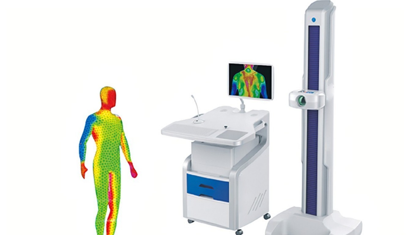
Medical thermal imaging equipment usually has two parts, an infrared thermal imaging camera, and a standard PC or laptop. These systems have only a few controls and are relatively easy to use.
Monitors are high-resolution full-color, isotherms, or grayscale, and typically include image processing, isothermal temperature mapping, and point-by-point temperature measurements using cursors or statistical regions of interest. The system measures temperatures from 10℃ to 55℃ with an accuracy of 0.1℃. Focus adjustments should cover a small area as small as 75 x 75mm.
These systems are PC-based and therefore capable of storing tens of thousands of images (and can be retrieved for later analysis). The ability to statistically analyze thermograms in the future is very important in clinical work. Copies of images can be easily sent (via email, floppy disk, etc.) to referring physicians or other healthcare professionals.
The medical applications of infrared thermal imaging are very broad, especially in the fields of rheumatology, neurology, oncology, physiotherapy, and sports medicine. Thermal imaging systems are an economical and easy-to-use tool for quickly and accurately examining and monitoring patients. The following are some applications of infrared thermal imaging in medical diagnosis.
Breast lesions
This is probably the area where medical thermal imaging has the most applications - breast cancer, benign tumors, mastitis, and fibrocystic breast disease.
The use of thermal imaging as a screening tool to detect breast cancer has been a hotly debated topic in the healthcare community over the past decade.
However, the technology has gained scientific acceptance, has been approved for screening purposes, and is clearly a powerful tool in the fight against breast cancer.
The concept is simple. Thermal imaging measures the heat from your body. The heat generated by metastatic cancer can be imaged with digital infrared imaging. This is due to two separate but interrelated factors.
The first is the metabolic activity of the tumor tissue compared to the temperature of the tissue adjacent to the tumor, in the opposite breast. Abnormal thermal signatures associated with tumor metabolism can be readily detected by comparing the problematic breasts to normal breasts that serve as the patient's own controls. These temperature differences are called Delta T.
The second method of detection is due to tumor angiogenesis.
That is, cancerous tumors produce a chemical that actually promotes the development of blood vessels that supply the area where the tumor is located. In infrared thermal imaging, normal blood vessels controlled by the sympathetic nervous system are essentially paralyzed, resulting in vasodilation or increased vessel size. Increased blood in this area simply means more heat due to angiogenesis combined with vasodilation, which can be recorded by thermal imaging procedures.
Because thermal imaging has been shown in many studies to be able to measure these thermal signatures years before conventional techniques can see a lump, and because the procedure does not use radiation, compresses breast tissue, and is completely safe, thermal imaging, or infrared thermal imaging, offers a safe Early warning detection system.
Extracranial vascular disease
In a similar way, various infrared thermal imaging related to blood flow through the vessels of the neck and head can be easily captured by thermal imaging. Because blood vessels in the face and skull pass through the very thin tissue between the skull bones and the skin that covers the skull, they are easily visualized with thermal imaging.
Because the vessels of the neck are very large-bore vessels, they are also easily visualized by thermography, and the possibility of developing vascular disease that can lead to stroke needs to be considered when doing thermography.
Use thermal imaging to differentiate between various types of headaches (migraine, cluster headache, cervical spine-related), facial nerve damage (such as a blow to the face or a car accident in which the face touches the windshield or steering wheel), TMJ disorders (temporomandibular joint) Visualization is a common aspect of head and neck thermography diagnosis and analysis.
Thermography's ability to safely display heat from jaws and teeth presents a very exciting opportunity to screen individuals for cavities and cavities without routine screening X-rays. In infrared thermal imaging, many patients have seen a thermal signature in the jaw associated with amalgam fillings, which can be toxic to specific patients. This field of thermal imaging is very promising.
Neuromusculoskeletal
This is one of the clearest examples of thermal imaging being able to accurately diagnose patients with a wide range of back, neck, and extremity infrared thermal imaging. In fact, it was in the late 1970s and 1980s that chiropractors, neurologists, and orthopedic surgeons used thermal imaging in cases of spinal injuries caused by car accidents and work-related injuries that really sparked clinical interest in this diagnostic tool. interest.
When muscle tissue is strained or torn, it releases chemicals that cause an increase in heat. This can be seen as a hyperthermic pattern in a muscle area or trigger point, as is the case with fibromyalgia. Thermal patterns were also seen on the legs and soles, suggesting changes in gait or weight-bearing mechanisms, which may be related to lower back or foot infrared thermal imaging.
In infrared thermal imaging, back strains produce very consistent thermal patterns that can tell us not only the likely source of spinal injury but also the areas of spinal compensation. In reality, a chiropractor may be treating the lower back, when the back or neck is actually the source of the problem.
Nerve injuries, such as herniated discs and compression of spinal nerve roots, show up on a thermogram in the exact opposite direction of muscle damage, by revealing areas of hypothermia in nerve bundles coming from the spine.
In this way, thermal imaging can demonstrate and document the permanence of spinal injuries that result in disability. This documentary, rather than diagnostic thermal imaging, has been used in trial courts for years to demonstrate injury and assist in the assessment of permanent damage.
Lower extremity vascular disease
The ability of thermal imaging to detect deep vein thrombosis and other circulatory disorders in the lower extremities is a very exciting application of this procedure as it allows us to painlessly and safely detect possible diseases that, if left unchecked, can lead to limb loss, or in some cases, increased likelihood of stroke.
Another largely overlooked aspect of thermal imaging is diabetic neuropathy in the foot that occurs before the foot becomes numb.
For example, we often see people with extremely cold feet on thermal imaging, even though they have no other symptoms.
Thermal imaging of the foot shows 1-2 degrees Celsius cooler than the calf, and usually, the toes are not visible in the camera because they become so cold. This can occur years before routine blood tests indicate diabetes, thus giving patients time to treat the infrared thermal imaging before permanent nerve damage occurs in the feet.
The above introduces the application of infrared thermal imaging in medical diagnosis. If you plan to buy an infrared thermal imaging camera, please contact us.
Javol is a professional custom infrared thermal imaging system supplier, we have a young and challenging technical research and development team. All product design engineers have bachelor's degrees or above. The team is personally led by senior doctors from famous universities to develop products that meet customer requirements. Everyone in the technical team has the expertise, including software design, hardware design, mechanical structure design, etc.
